3Rs implementation
|
The first R requires asking, as a preliminary step, whether it is possible to replace the chosen animal model with methods that allow the same result to be achieved without using animals (Replacement, or Replacement), or by using a “simpler” animal model (partial replacement). If this is impossible, every means must be used to minimize the number of individuals employed, without compromising the reliability of the result (Reduction, Reduction). Finally, the animals used – regardless of species – must be treated using the most appropriate measures to make procedures less impactful on their welfare, reducing as much as possible any type of suffering that may derive from experimental procedures or housing conditions (Refinement, Refinement). The “3Rs” have evolved over time, even though their underlying principles have remained the same, and they are now formally integrated into European and national legislation protecting the welfare of animals used for scientific purposes. |
 |
|
Replacement The ongoing basic research projects aim to understand the neural mechanisms underlying high-level motor and socio-cognitive functions, expressed in the behavioural repertoire of only a few primate species in addition to Humans. The first and most obvious option to replace the animal model would be to study the same functions directly in human participants. This is indeed possible and widely pursued thanks to indirect and non-invasive techniques for investigating brain activity, such as functional magnetic resonance imaging (fMRI), high-density electroencephalography (EEG), transcranial magnetic stimulation (TMS) and magnetoencephalography (MEG). In addition, over the last 20 years, the use of prolonged intracranial electroencephalographic monitoring for pre-surgical evaluation of epileptogenic foci in patients with drug-resistant epilepsy and negative MRI findings has made it possible to further articulate the study of human brain functions with invasive techniques used primarily for clinical purposes (see for example1). Computational simulation models offer an additional possibility to integrate acquired data and formulate empirically grounded hypotheses and predictions, but they still require direct experimental data. These approaches have contributed to a marked reduction in the use of animals – in particular non-human primates – employed in neuroscientific research (from ~8,000 to ~6,000 in Europe between 2008 and 2011, −25%, source: SCHEER report 2017). However, none of these techniques can currently replace the animal model for studies at the single-neuron and neuronal-network level, which require direct electrophysiological recordings. Studies with simultaneous recordings from multiple regions are essential to link the single-cell level to the macrosystem of neuronal networks, as well as to enable causal inferences about the mechanisms generating behaviour. This paradigm, already widely developed in murine models2,3, cannot be transferred to our research questions when fine motor repertoire, communication and social interaction require anatomo-functional and behavioural homologies that only non-human primates provide. Within this framework, macaques of the genus Macaca represent the species with the lowest neurological development that can be used to achieve the objectives of the projects. |
||
|
Macaques of the genus Macaca (fascicularis and mulatta) are reference species in biomedical and neuroscientific research4, with anatomo-functional, cognitive and behavioural homologies necessary and sufficient for results that are relevant on a comparative and evolutionary level5. The motor skills of the limbs and the fine use of the hands to reach and manipulate objects, for example, rely on evolutionarily conserved mechanisms and substrates6. Macaques display a broad social and affiliative repertoire, expressed through postures, facial gestures, vocalizations and multisensory combinations, which is not comparable to species with lower neurological development7. |
||
|
|
|
|
|
More specifically, the species Macaca mulatta is widely used in behavioural neurophysiology studies for its excellent adaptability and tolerance to housing and laboratory conditions, in compliance with adequate environmental enrichment measures and social housing8. It is also the species with which our group has the greatest direct experience: biology and ethology have been investigated in numerous neurophysiological research projects at the University of Parma, authorized by the Ministry of Health, and consolidated through specific training for personnel performing the tasks and functions referred to in art. 23, paragraph 2, Legislative Decree 26/2014 (DM 5 August 2021, specific module for non-human primates) and ongoing collaborations with other primatology centres and research groups in Europe and worldwide (see Collaborations). |
||
|
Reduction In neuroscientific studies on non-human primates, within-subject designs are predominantly adopted: sample size is defined not by the number of animals, but by the number of independent measurements obtained reliably and reproducibly in the individual subject. To address inter-individual variability and ensure data robustness and reproducibility, neurophysiological literature requires as a standard the replication of the effect in a second animal, although a substantial number of recordings and observations obtained in a single subject is increasingly considered useful and relevant9. Consequently, the minimum number of animals necessary and sufficient for each neurophysiological experiment is typically 2. The use of new technologies that allow the same animals to be used in different experiments (e.g., injections of pharmacologically active substances and neuroanatomical studies in the same subject) makes it possible to increase the quality and completeness of results and, at the same time, reduce the overall number of animals needed for an equivalent amount of information, without an increase in “cumulative severity”, i.e., without causing greater harm or suffering to the individual animal.
Refinement in non-human primate research is both an ethical and regulatory obligation and an essential means to optimize animals’ cooperation and maximize data quality (see Refinement techniques in non-human primate neuroscientific research). All procedures – from housing to surgical interventions and experimental sessions – are set according to the best international standards. Our effort to definitively move beyond head-fixation systems in favour of wireless technologies with freely moving animals represents a qualitative leap that goes beyond current standards and embraces the most recent European recommendations (cf. SCHEER report 2017). Animals are sourced from authorized European suppliers or, preferably, European breeding centres able to guarantee the availability of so-called second-generation, or F2, animals, meaning that neither the animals nor their parents are ever directly taken from the wild. Transport is carried out by trained and authorized personnel using suitable vehicles. From acquisition onwards, with the aim of improving the “cumulative life experience”, we apply refinement measures that involve all management aspects, from housing to experimental procedures, as detailed in the following sections.
▪ Training of all personnel. Assessing, monitoring and optimizing the environment and procedures throughout the animals’ entire life cycle can bring substantial benefits only insofar as they are carried out by expert and adequately trained staff. For this reason, all personnel with direct responsibility for procedures or daily animal management receive appropriate specific theoretical and practical training, demonstrated by completion of the training required for personnel performing the tasks and functions referred to in art. 23, paragraph 2, of Legislative Decree no. 26/2014 (DM 5 August 2021), specific for non-human primates used for experimental purposes, before starting any interaction with the animals. Continuous exchange with staff (colleagues and veterinarians) working in other European primatology centres is a source of ongoing updating and continuous training, in addition to the periodic updating of staff obtained through participation in seminars and specific events on 3Rs and animal welfare. |
||
|
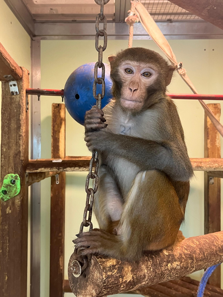
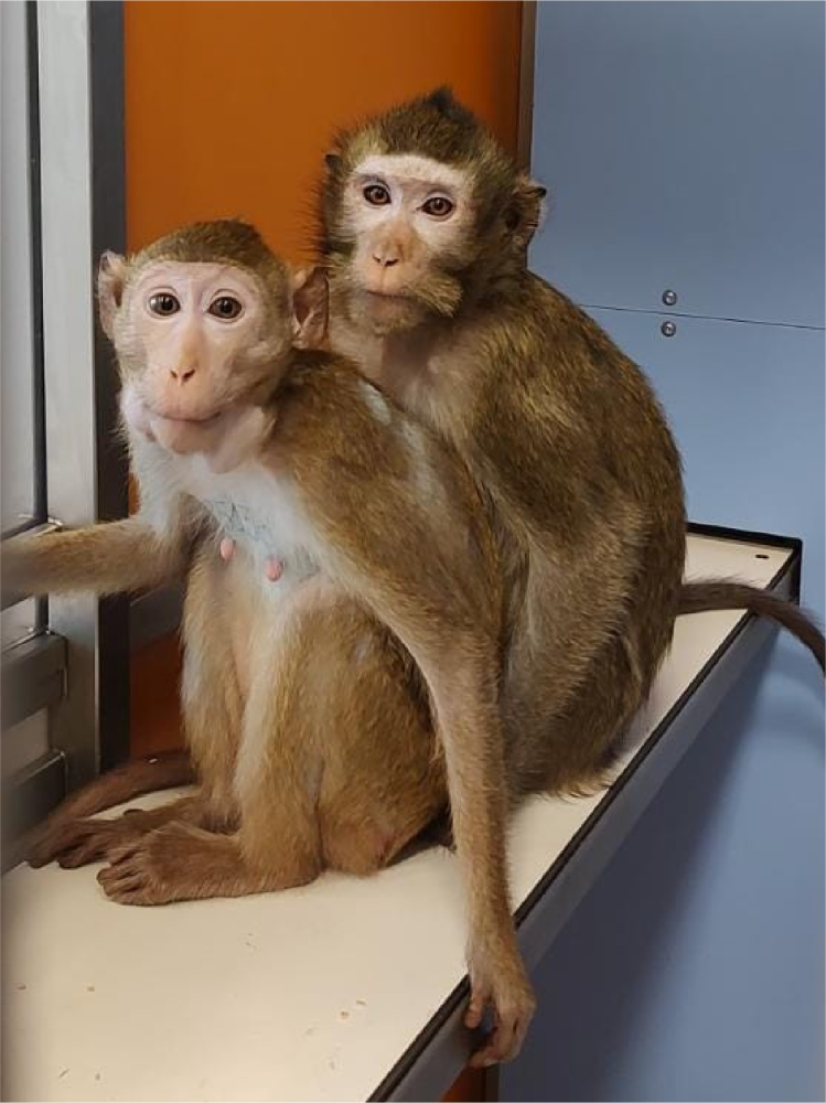
▪ Housing and environmental enrichment. Our animal facility adopts a communicating-cage system compliant with Directive 2010/63/EU and Legislative Decree 26/2014, designed and built by a leading company in the sector in collaboration with the main European primatology centres. Animals (up to a maximum of 20) are housed in pairs or small groups in spaces always above legal limits (≥ 2.5 m3 vs 1.8 m3 required per animal). Environmental enrichment follows a daily rotation program that includes physical/structural interventions (vines, hanging toys, climbing elements), sensory enrichment (mirrors, visual and/or auditory stimuli), food-based enrichment (sawdust or natural bark substrates for scattering seeds and small foods; perforated objects for “foraging”), cognitive-occupational enrichment (puzzles and problem-solving activities), and social enrichment. Recreation cages with wooden structures and swings are also available, accessible to pairs or small groups of animals on a daily rotation, allowing them to maintain and exercise their full species-specific behavioural repertoire (e.g., climbing, jumping, exploration). Within the animal facility, there is also a housing room for groups of animals (L × W × H: 3.43 × 2.3 × 2.72 m), equipped with a large wooden structure for climbing and jumping, natural bark flooring, and direct connection to other cages via an elevated tunnel. A sound diffusion system and video monitors provide additional sensory and cognitive enrichment. The environment benefits from large windows for optimal natural lighting and maintenance of regular day/night cycles; artificial lighting is controlled by timers. Climate is managed by a temperature control system with 24/7 active alarms. Hygienic-sanitary management of the facilities and daily nutrition are handled by trained technical staff operating according to indications and under the supervision of research staff and technical research-support personnel. |
|
|||||
|
▪ Training with positive reinforcement. All animals are trained exclusively using methods based on positive reinforcement and voluntary cooperation. The pathway begins with a phase of progressive familiarization with experimenters and equipment, during which simple commands are given through the cage and reinforced with small food rewards (e.g., fruit), distinct from the daily dry ration. Habituation and specific training procedures are performed by competent personnel using operant conditioning and positive reinforcement techniques10. Clicker training is used systematically as an immediate auditory signal that marks the correct behaviour and precedes the reward, facilitating and accelerating learning11,12. For experimental sessions, animals are progressively trained to approach, enter and remain in the primate chair for initially short periods that gradually increase, without the use of collars or rigid spacers13,14. Sessions are planned with gradual goals, kept to a limited duration, and are interrupted or modulated in the presence of stress signals, to safeguard animal welfare and data quality. In our laboratory, we are systematically adopting procedures that eliminate head-fixation devices and implement the use of transparent plexiglass carriers for transferring animals to the environment equipped for wireless recordings. This approach enables shorter habituation pathways (generally a few weeks) with minimal signs of stress or refusal compared to traditional protocols, which may require up to a year, particularly for the delicate phase of habituating animals to repeatedly accept limited mobility in the primate chair. Graduality remains a core principle: training proceeds step by step, thereby reducing the risk of regressions and promoting voluntary cooperation. Specific training to experimental tasks, when necessary, is conducted with the same evidence-based behavioural methodologies: task analysis, breaking down behaviour into elementary components, differential reinforcement of spontaneous behaviours that approach the target, response shaping and fading-out of cues used as intermediate steps. The goal is to select and reinforce behaviours closest to the desired behaviour, facilitating extinction of alternative behaviours and leading to the progressive acquisition of the skills required even in cognitively complex tasks. This pathway, fully in line with the practices of the best dog trainers, represents an advancement within the 3R landscape, combining animal welfare and data quality. |
||||||
|
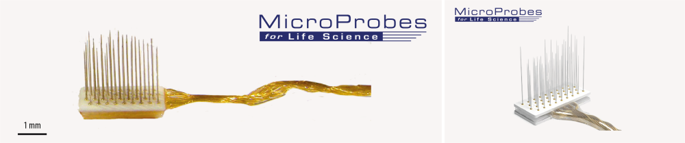
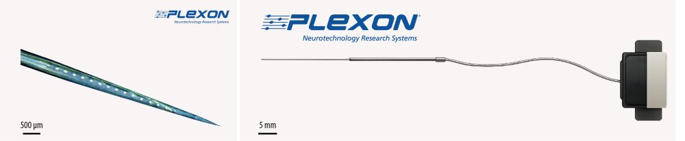
▪ Miniaturization and refinement of invasive devices. All implanted devices (such as the interconnection system for wireless transmitters) are made of biocompatible materials and reduced to the smallest possible size. The multielectrode probes used typically have diameters between 50–100 µm, thus limiting tissue damage to negligible levels, as confirmed by histological analyses in previous studies15. This also allows animals to be returned to their group at the end of the experiment without the need for euthanasia unless required by the experimental protocol to allow reconstruction of the anatomy and connections of the brain regions studied. |
|
|||||
|
Head-fixation devices still in use, employed only where necessary, have proven to be particularly well tolerated, showing excellent osseointegration also evidenced by post-mortem examinations16. In line with the refinement principle (3R), we are moving steadily towards a non-invasive head immobilization system, completely external and temporary, which does not involve any fixed cranial implant. This support is applied only for a few moments, exclusively to stabilize the head while connecting the wireless recording system, and then removed. The approach reduces animal handling, shortens pre-session preparation, and eliminates the risk of infectious or inflammatory complications associated with a chronic implant. In parallel, when possible, we adopt chronic or semi-chronic recordings with implants performed on intact cranial bone in the region of interest (still carried out under general anaesthesia), minimizing handling before each session and drastically reducing the risks of infection or inflammation, thus further contributing to improved animal welfare. |
||||||
|
▪ Anaesthetics, surgical techniques and post-operative pharmacological treatment. All major surgical procedures are performed under general gaseous anaesthesia with halogenated agents administered and controlled by a veterinarian with specific training for personnel performing the tasks and functions referred to in art. 23, paragraph 2, Legislative Decree 26/2014 (DM 5 August 2021, specific module for non-human primates) and experience in anaesthesia of non-human primates. Post-operative recovery is followed by the personnel responsible for animal management under the supervision of the Designated Veterinarian and the anaesthetist veterinarian, who prescribe and adjust antibiotic, anti-inflammatory and analgesic therapy until full recovery. |
 |
|||||
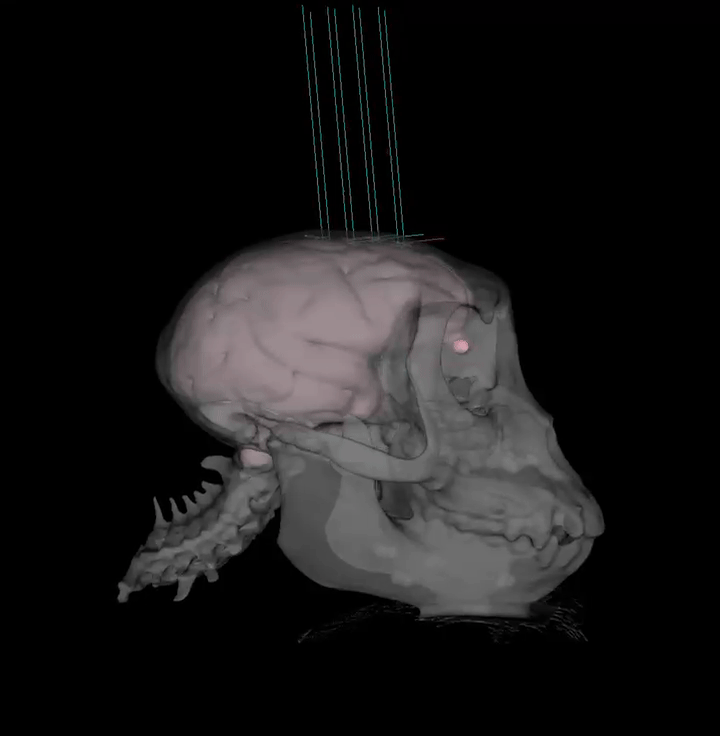
| ▪ Recording techniques that maximize the quantity and quality of data, reducing the number and duration of sessions. A further refinement element consists in optimizing the use of MRI and CT under anaesthesia to obtain high-resolution images of brain and skull morphology. The 3D reconstruction of a plastic model of each animal’s skull makes it possible to anatomically conform the devices to be implanted to the specific structure of the bone: results obtained so far show lighter and less invasive implants, which do not require particular disinfection or cleaning procedures after implantation.
Studying brain morphology allows precise identification of the region of interest and is the basis for the use of chronic or semi-chronic probes implanted precisely in the target region. Multichannel recording makes it possible to acquire activity from hundreds of sites simultaneously, thus limiting tissue damage and maximizing the quantity and quality of data. Monitoring behavioural parameters, such as movement kinematics or eye position, is performed with totally non-invasive methodologies that ensure signal stability under any visible light condition. |
|
| ▪ Use of the same animals for neurophysiological and neuroanatomical studies. In our laboratory, the same animals can be employed for neurophysiological studies and, subsequently, neuroanatomical studies when the objectives of the study require it and, in that case, without an increase in cumulative severity or in the number of animals needed. Implantation of intracortical probes is performed using miniaturized devices and does not in itself cause relevant tissue damage or suffering for the animal. When the goal is to define the connectivity of regions investigated electrophysiologically, we use injectrodes, i.e., silicon probes equipped with one or more microchannels for the infusion of microlitres of pharmacologically active substances or, at the end of experiments, neuronal tracers. These tracers, with retrograde or anterograde transport, allow – after an adequate survival interval of the animal that depends on the transport speed of the specific tracer – post-mortem verification of the anatomical connectivity of the same region studied functionally.
Neuroanatomical studies do not require additional procedures beyond those already planned for neurophysiological studies and do not require dedicated animals; consequently, they do not produce additive effects in terms of “cumulative severity”. When it is scientifically necessary to precisely reconstruct the anatomical network of the studied brain, painless euthanasia is performed according to the methods provided for the species. When such verification is not required or is not scientifically necessary as part of the experimental plan, implants are surgically removed and animals are included in a relocation program at accredited rescue centres, in compliance with current regulations and following veterinary assessment (health suitability, observation period, behavioural requirements). |
|
| ▪ Continuous monitoring of animal welfare. Despite the measures described, unpredictable adverse events may still occur, related or unrelated to experimental procedures or housing conditions, which can affect the animal’s welfare (e.g., accidental trauma, wounds from conflict with a partner, transient infections or organ dysfunctions attributable or not to experimental procedures). Such events may also occur independently of research activity, as happens for any domestic animal.
To prevent and effectively manage possible adverse events, we adopt continuous welfare monitoring carried out by trained and qualified personnel. For each subject, an individual clinical assessment form is compiled upon entry into the animal facility and updated daily throughout the entire life cycle. Daily monitoring records broader information than that required by the personal history file provided by regulations: clinical and behavioural observations, amount and type of food and liquids consumed, body weight, notable events or behaviours, type of enrichment provided and degree of interaction, performance during training and any experimental tasks. All animals involved in any procedure, even only during training, are observed by – or interact every day with – at least one reference experimenter and/or technician, able to promptly detect any deviation from the optimal state and activate appropriate corrective actions such as veterinary assessment, modification or pause in training, adaptation of environmental conditions or enrichment. The collected information allows protocols to be modulated and optimized based on the individual characteristics of each animal. |
References
[1] F. Caruana et al., “Decomposing Tool-Action Observation: A Stereo-EEG Study,” Cereb. Cortex, vol. 27, no. 8, pp. 4229–4243, Aug. 2017.
[2] G. Buzsáki et al., “Tools for Probing Local Circuits: High-Density Silicon Probes Combined with Optogenetics,” Neuron, vol. 86, no. 1, pp. 92–105, 2015.
[3] P. Tovote, J. P. Fadok, and A. Lüthi, “Neuronal circuits for fear and anxiety,” Nat. Rev. Neurosci., vol. 16, p. 317, May 2015.
[4] P. K. A. et al., “Why primate models matter,” Am. J. Primatol., vol. 76, no. 9, pp. 801–827, Aug. 2014.
[5] K. J. H., “The evolution of brains from early mammals to humans,” Wiley Interdiscip. Rev. Cogn. Sci., vol. 4, no. 1, pp. 33–45, Nov. 2012.
[6] E. Borra, M. Gerbella, S. Rozzi, and G. Luppino, “The macaque lateral grasping network: A neural substrate for generating purposeful hand actions,” Neurosci. Biobehav. Rev., vol. 75, pp. 65–90, 2017.
[7] J. Fooden, “Systematic review of the rhesus macaque, Macaca mulatta (Zimmermann, 1780).,” FieldianaZoology, vol. 96, pp. 1–180, 2000.
[8] H. D. L., B. Eliza, V. Jessica, M. Brenda, and C. John, “Laboratory rhesus macaque social housing and social changes: Implications for research,” Am. J. Primatol., vol. 79, no. 1, p. e22528, Dec. 2016.
[9] P. Fries, E. Maris, "What to Do If N Is Two?", J Cogn Neurosci 2022; 34 (7): 1114–1118.
[10] G. E. Laule, M. A. Bloomsmith, and S. J. Schapiro, “The Use of Positive Reinforcement Training Techniques to Enhance the Care, Management, and Welfare of Primates in the Laboratory,” J. Appl. Anim. Welf. Sci., vol. 6, no. 3, pp. 163–173, Jul. 2003.
[11] S. J. Schapiro, M. A. Bloomsmith, and G. E. Laule, “Positive reinforcement training as a technique to alter nonhuman primate behavior: Quantitative assessments of effectiveness,” Journal of Applied Animal Welfare Science, vol. 6, no. 3. pp. 175–187, 2003.
[12] A. L. Fernström, H. Fredlund, M. Spångberg, and K. Westlund, “Positive reinforcement training in rhesus macaques-training progress as a result of training frequency,” Am. J. Primatol., vol. 71, no. 5, pp. 373–379, 2009.
[13] L. Scott, P. Pearce, S. Fairhall, N. Muggleton, and J. Smith, “Training nonhuman primates to cooperate with scientific procedures in applied biomedical research,” Journal of Applied Animal Welfare Science, vol. 6, no. 3. pp. 199–207, 2003.
[14] S. Mason, E. Premereur, V. Pelekanos, A. Emberton, P. Honess, AS. Mitchell, "Effective chair training methods for neuroscience research involving rhesus macaques (Macaca mulatta)", J Neurosci Methods, 2019.
[15] F. Barz et al., “Versatile, modular 3D microelectrode arrays for neuronal ensemble recordings: From design to fabrication, assembly, and functional validation in non-human primates,” J. Neural Eng., vol. 14, no. 3, 2017.
[16] A. Kohn, “Visual Adaptation: Physiology, Mechanisms, and Functional Benefits,” J. Neurophysiol., vol. 97, no. 5, pp. 3155–3164, May 2007.